


Introduction
- Overview of Digital Imaging in Dentistry: Digital imaging has revolutionized the way dental professionals diagnose and treat oral health conditions. These technologies enhance the precision, speed, and efficiency of dental procedures.
- Significance of Accuracy in Dental Diagnosis: Accurate diagnosis is crucial for determining the right treatment plan and avoiding unnecessary procedures, saving time and resources for both patients and dentists.
- Purpose of the Article: This article will explore the role of digital imaging in improving dental diagnosis and treatment, focusing on its benefits, applications, and how it leads to better patient outcomes.
1. What Is Digital Imaging in Dentistry?
- Definition of Digital Imaging: Digital imaging refers to the use of advanced technology to capture high-quality images of a patient’s teeth, gums, and jaw. Unlike traditional X-rays, digital images are captured electronically, allowing for instant viewing and enhanced visualization.
- Types of Digital Imaging in Dentistry:
- Intraoral Imaging: Small sensors placed inside the patient’s mouth to capture detailed images of individual teeth.
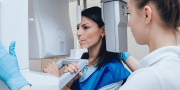
- Extraoral Imaging: Images taken from outside the mouth, such as panoramic X-rays that show the entire jaw.
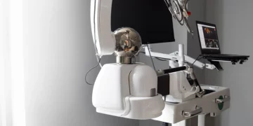
- Cone Beam Computed Tomography (CBCT): A 3D imaging technology that allows for a more detailed view of the teeth, soft tissues, and bone structures.
- How Digital Imaging Differs from Traditional Methods: Traditional X-rays use film to capture images, while digital imaging uses electronic sensors, resulting in faster processing times, clearer images, and lower radiation exposure.
2. Benefits of Digital Imaging in Dentistry
- Enhanced Accuracy of Diagnosis:
- Digital imaging allows for high-resolution, clear images that provide dentists with a better view of the teeth and gums, making it easier to detect issues like cavities, gum disease, fractures, and tumors.
- Dentists can view images immediately, ensuring prompt diagnoses and reducing the need for retakes.
- Reduced Radiation Exposure:
- Digital X-rays emit much lower levels of radiation compared to traditional film-based X-rays, making them safer for both patients and dental professionals.
- The ability to take multiple images with lower radiation levels improves patient safety, especially for those who require frequent imaging (e.g., children, pregnant women).
- Faster Image Processing:
- Digital images are immediately available for review, meaning that treatment decisions can be made quickly, reducing patient wait times and the overall duration of dental visits.
- Unlike traditional X-rays, digital images do not need to be developed in a darkroom, eliminating wait times for the processing of film.
- Increased Comfort for Patients:
- Digital sensors are often smaller and more comfortable than traditional film-based X-ray equipment, reducing discomfort for patients during the procedure.
- The lack of chemicals used in the imaging process also contributes to a more comfortable environment.
3. Applications of Digital Imaging in Dental Diagnosis
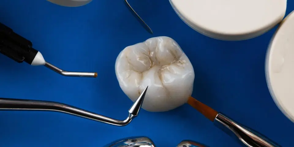
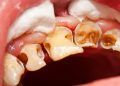
- Detecting Cavities and Tooth Decay:
- Digital X-rays help dentists detect cavities at the earliest stages, even before they are visible to the naked eye. This allows for early intervention and less invasive treatments.
- Identifying Gum Disease:
- Images of the gums can show signs of inflammation, infection, or bone loss, which are early indicators of gum disease such as gingivitis and periodontitis.
- Regular digital imaging can track the progression of gum disease and aid in designing more effective treatment plans.
- Assessing Bone Loss and Jaw Health:
- CBCT and panoramic X-rays provide an excellent view of the jaw and bone structures, allowing dentists to identify issues such as bone loss, cysts, and tumors.
- These images can be used to determine if bone grafting or other surgical interventions are necessary for a patient with advanced gum disease or injury.
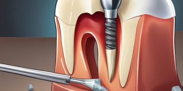
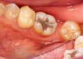
- Planning for Implants and Other Restorative Procedures:
- For dental implant procedures, digital imaging is essential for accurate placement. CBCT imaging provides detailed 3D views of the bone structure, ensuring implants are placed correctly and in a way that promotes long-term success.
- Monitoring Treatment Progress:
- Digital imaging allows for continuous monitoring of how treatment plans are progressing, helping dentists evaluate the effectiveness of treatments like orthodontics or periodontal therapy.
4. How Digital Imaging Improves Treatment Planning
- Improved Visualization for Better Planning:
- High-quality, detailed images enable dentists to visualize a patient’s mouth more comprehensively. This can lead to better treatment planning, as the dentist can identify potential complications or hidden issues that might affect treatment success.
- 3D Imaging for Complex Cases:
- With 3D imaging (such as CBCT), dentists can plan complex procedures, such as implant placement, root canals, or jaw surgery with greater accuracy, minimizing the risk of errors.
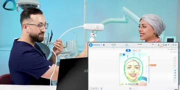
- Predicting Treatment Outcomes:
- Digital imaging software can be used to simulate treatment outcomes, such as how teeth will align after braces or how a tooth will look after restorative work. This helps both the patient and dentist visualize the final result, ensuring alignment with patient expectations.
5. How Digital Imaging Aids in Early Detection and Prevention
- Cavity Detection at an Early Stage:
- Early detection of cavities allows for preventative treatments like fluoride application, which can prevent cavities from progressing to the point where they require invasive treatment such as fillings.
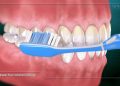
- Gum Disease Prevention:
- Regular digital imaging helps identify early stages of gum disease, allowing dentists to recommend preventive treatments (e.g., deep cleaning, scaling, and root planing) to stop the disease before it becomes more severe.
- Tracking Changes Over Time:
- Digital images can be stored and compared over time, providing a clear picture of how a patient’s oral health is changing. This long-term tracking can help detect problems before they become serious.
- Preventive Care for High-Risk Patients:
- For patients at high risk of oral health issues (e.g., individuals with diabetes or a history of cavities), digital imaging provides a valuable tool for more frequent monitoring and proactive care.
6. The Role of Digital Imaging in Orthodontics
- Planning and Monitoring Braces and Aligners:
- Digital imaging is used in orthodontics to plan the movement of teeth during braces treatment. The images help the orthodontist predict how teeth will shift over time.
- Tracking Treatment Progress:
- Digital X-rays and 3D scans help orthodontists track how the treatment is progressing, and whether any adjustments are needed. It also helps in monitoring bone growth and ensuring that teeth are moving correctly.
- Minimizing Treatment Time:
- Digital imaging helps to reduce errors, streamline the treatment process, and shorten the time spent in braces. The precise planning made possible through digital imaging leads to more effective treatment and quicker results.
7. Patient Education and Communication Through Digital Imaging
- Better Patient Understanding:
- Digital images provide clear visuals that help patients understand their dental conditions. A patient is more likely to follow a recommended treatment plan when they can see and understand the problem.
- Improved Communication Between Patients and Dentists:
- Digital images allow dentists to explain diagnoses and treatment plans in a visual, easy-to-understand manner. Patients can be shown the areas of concern and the planned interventions.
- Increasing Patient Confidence:
- By showing patients their oral health status through digital images, dentists can increase trust and make patients feel more confident in the proposed treatments.
8. Digital Imaging and Its Impact on Treatment Outcomes
- Improved Treatment Success Rates:
- The enhanced accuracy of digital imaging ensures that treatments are planned and executed with more precision, leading to higher success rates, especially in complex procedures such as implants or orthodontic work.
- Minimizing Treatment Complications:
- By detecting problems early and accurately, digital imaging helps avoid complications during or after treatment. For example, accurate root canal imaging can prevent damage to surrounding tissues and nerves.
9. The Future of Digital Imaging in Dentistry
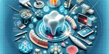
- Advances in Imaging Technology:
- As technology continues to advance, future developments in digital imaging could include even higher resolution images, faster processing, and even more accurate 3D scans.
- Integration with Artificial Intelligence (AI):
- AI can be integrated with digital imaging to automate certain diagnostic tasks, such as detecting cavities or gum disease. AI could also assist in treatment planning by analyzing vast amounts of data to suggest the most effective treatments.
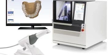
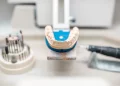
- Expansion of Intraoral Scanning:
- Intraoral scanning, where a 3D scan of the mouth is taken, is becoming more common and may replace traditional molds used in procedures like crowns and bridges.
- Telemedicine and Remote Diagnosis:
- Digital images can be shared easily, opening the door to telemedicine, where dentists can remotely review images and offer consultations to patients in underserved areas.
10. Conclusion
- Summary of Benefits:
- Digital imaging significantly improves the accuracy and efficiency of dental diagnoses and treatments. It offers better image quality, reduces radiation exposure, speeds up the diagnostic process, and enhances treatment planning.
- Final Thoughts:
- As digital imaging continues to evolve, its role in dental care will become even more integral to providing personalized, accurate, and effective treatments that enhance patient outcomes.


Discussion about this post