


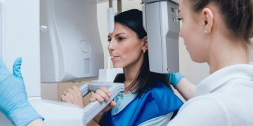


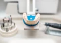
Dental X-rays have always been an essential tool for diagnosing problems that cannot be seen with the naked eye—cavities between teeth, bone loss, impacted teeth, infections, and early developmental issues. But for decades, patients felt uneasy about the word “X-ray,” largely due to concerns about radiation and outdated imaging methods. Fortunately, the evolution of digital X-ray technology has transformed this experience completely. Today’s digital systems expose patients to far less radiation, provide sharper images, deliver instant results, and integrate seamlessly with cutting-edge diagnostic tools.
In this article, we explore how digital X-rays have progressed, why they are significantly safer than traditional film X-rays, and how these improvements empower both patients and dental professionals.
1. How Has Radiation Exposure Been Reduced in Modern Digital X-Rays?
One of the biggest advancements in dental imaging is the dramatic reduction in radiation. For many patients, this improvement removes one of the main barriers to feeling comfortable with X-ray diagnostics.
Digital sensors require far less radiation
Traditional film X-rays used photographic plates that needed high radiation levels to activate light-sensitive chemicals. By contrast, digital sensors are:
- More sensitive to light
- Capable of capturing detailed images with minimal exposure
- Designed to maximize usable information from very low signals
As a result, digital radiography reduces radiation by up to 80–90% compared with conventional film.
More focused radiation beams
Modern X-ray machines use:
- Collimators that narrow the beam
- Pulse-based radiation emissions
- Sharper aim to limit scatter
All these enhancements minimize overall exposure.
Faster image capture means shorter exposure time
Because digital sensors capture data instantly, there’s no need for prolonged radiation or repeated shots.
Protective standards continue to improve
Newer machines follow:
- ALARA guidelines (As Low As Reasonably Achievable)
- Updated filtration standards
- Shielding technologies
- Better calibration controls
Altogether, modern digital X-rays offer some of the safest imaging available in healthcare—often lower in radiation than a short airplane flight.
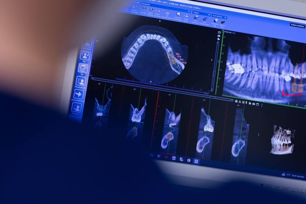
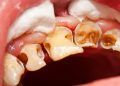
2. Why Do Digital X-Rays Offer Higher Clarity and More Diagnostic Detail?
Clearer images lead to more accurate diagnoses, earlier detection, and better treatment planning. Digital radiography provides dentists with unprecedented image quality.
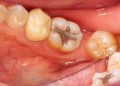
High-resolution sensors capture micro-details
Digital sensors can capture:
- Microfractures
- Early decay
- Subtle bone density changes
- Periodontal pocket patterns
- Hidden infections
Film X-rays simply cannot record this level of detail consistently.
Image enhancement tools improve visibility
Dentists can adjust:
- Brightness
- Contrast
- Magnification
- Sharpness
- Zoom
- Filters
This reduces guesswork and eliminates the errors caused by film overexposure or underexposure.
Consistent results without chemical processing
Film X-rays depended on proper development techniques. Factors like room temperature, chemical freshness, and technician skill impacted clarity. Digital imaging eliminates all of these variables, ensuring precise, predictable images every time.
Better visualization leads to earlier intervention
Higher clarity helps dentists catch problems while they are still small and reversible—reducing the need for invasive treatment later on.
3. How Do Digital X-Rays Enable Faster and More Accurate Diagnosis?
Speed is one of the most transformative benefits of digital radiography. What once took minutes or hours now takes seconds.
Instant image display
Digital X-rays appear on the screen immediately. No waiting. No retakes due to poor development. This:
- Speeds up appointments
- Allows immediate diagnosis
- Enables real-time treatment planning
Better communication with patients
Dentists can show patients high-resolution images on large screens, making it easier to explain:
- Decay
- Bone loss
- Infection
- Tooth fractures
Patients understand their condition more clearly and make more informed treatment decisions.
Digital storage and sharing
Images can be:
- Emailed
- Saved
- Shared with specialists
- Added to patient records
This dramatically improves care coordination, especially for orthodontics, periodontics, oral surgery, and endodontics.
Fewer retakes improve safety and efficiency
Because the capture process is so sensitive, retakes are rare—further reducing radiation exposure and saving time.
4. What Benefits Do Patients Experience Directly from Digital Dental X-Rays?
Beyond the technical improvements, digital X-rays create a far more comfortable and reassuring experience for patients.
Lower exposure reduces anxiety
Patients concerned about radiation—children, pregnant individuals, or medically compromised patients—benefit from safer imaging.
More comfortable sensors
New sensor designs are:
- Thinner
- Smaller
- More ergonomic
- Less rigid
This reduces discomfort and gag reflex sensitivity.
Faster appointments
Shorter imaging times reduce chair time, making dental visits more efficient and less stressful.
Greater transparency and trust
Dentists can walk patients through their images in real time, allowing people to “see what the dentist sees.” This visual understanding improves:
- Treatment acceptance
- Patient confidence
- Oral health awareness
Better long-term outcomes
Earlier detection means smaller fillings, fewer root canals, less tooth loss, and lower treatment costs overall.
5. What Does the Next Generation of Dental Imaging Look Like?
Digital X-rays continue to evolve rapidly, and the next decade promises even more breakthroughs.
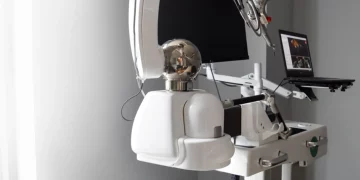
3D CBCT (Cone Beam Computed Tomography)
CBCT systems create three-dimensional models of the:
- Jaw
- Sinuses
- Nerves
- Bone structure
This is vital for:
- Implant planning
- Root canal treatment
- Orthodontics
- Trauma assessment
Future CBCT units will offer:
- Even lower radiation
- Higher precision
- Faster scanning
AI-assisted diagnostics
Artificial intelligence is revolutionizing dentistry by:
- Detecting cavities automatically
- Measuring bone density
- Predicting treatment outcomes
- Highlighting abnormalities
AI improves accuracy and reduces human error.
Ultralow-dose imaging protocols
New sensors aim to cut radiation exposure even further, approaching levels close to background environmental exposure.
Portable and handheld X-ray systems
Lightweight devices will make imaging easier in:
- Remote areas
- Hospitals
- Mobile dental units
- Nursing homes
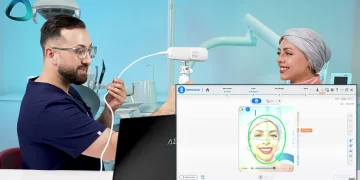
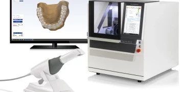
Integration with digital smile design and treatment planning
Future systems will merge:
- X-rays
- 3D models
- Intraoral scans
- Facial imaging
This unified digital workflow represents the next era of precision dentistry.
Final Thoughts: Why Are Digital X-Rays the Modern Standard?
Digital X-rays have redefined safety, clarity, efficiency, and patient-centered care in dentistry. With dramatically lower radiation, superior image quality, instant diagnostics, and innovations like AI and 3D imaging on the horizon, digital radiography is no longer just an upgrade—it is the foundation for modern dental diagnosis and treatment.
As technology continues to evolve, patients can expect even safer, more comfortable, and more precise imaging experiences in the years ahead.


Discussion about this post